Interstitial Fluid Dynamics, Lymphatic Drainage, and Plasma Refill
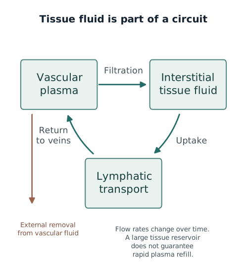
Edema tells us that fluid has accumulated in tissue. It does not tell us how quickly that fluid can return to plasma. To understand both accumulation and clearance, follow fluid across the microvascular wall, through the tissue matrix, into lymphatics, and back to the circulation.
18.1 The Interstitium Is a Structured Fluid Compartment
The interstitium is the space between tissue cells and the blood and lymphatic vessels around them. It contains water, dissolved substances, proteins, resident cells, and extracellular matrix. Fluid must move through this structure to reach cells or drainage pathways. It is not simply held in an empty chamber.
Collagen fibers help support the tissue. Proteoglycans and glycosaminoglycans form a hydrated matrix between those fibers. The arrangement of this matrix influences how readily water moves. A tissue can contain substantial water without allowing it to flow as freely as saline through a tube.
Pressure differences drive interstitial fluid movement, while tissue structure resists it. Hydraulic conductivity describes how easily fluid moves through a material under a pressure gradient. It is distinct from compliance, which describes how much tissue volume changes when pressure changes. A tissue that expands easily does not necessarily drain quickly.
Guyton and colleagues measured fluid mobility in dog subcutaneous tissue. Resistance changed with interstitial pressure, supporting the importance of tissue mechanics as well as fluid volume. The relationship measured in that tissue cannot be assigned unchanged to the kidney, lung, or another organ. 1
As loose tissue swells, expanded spaces can permit fluid to move more readily. Pressure from a finger can displace some of this fluid, producing pitting. The indentation shows that the tissue can deform and fluid can redistribute locally. It does not measure how rapidly fluid can enter lymphatics or refill plasma.
Location also determines the consequences of accumulation. Loose subcutaneous tissue can expand considerably. A kidney capsule or a fascial boundary restricts expansion. In a constrained space, a smaller volume increase may raise surrounding pressure enough to compress small vessels. Water between capillaries and cells can also lengthen diffusion paths. The effect of edema therefore depends on its location and mechanical behavior, as well as its volume.
18.2 Interstitial Pressure and Compliance Change with Swelling
Interstitial hydrostatic pressure is the pressure within tissue fluid. A negative value means pressure below the atmospheric reference, not absence of fluid. Some normally hydrated loose tissues have slightly negative pressures. Other tissues have different pressures because their structure, boundaries, and surrounding forces differ.
Compliance describes how much extra fluid volume accompanies a pressure increase. A highly compliant tissue accommodates a relatively large volume change with a small pressure change. A poorly compliant tissue develops a larger pressure rise for the same added volume.
The numerator is the added or lost tissue-fluid volume. The denominator is the accompanying pressure change. If volume is measured in mL and pressure in mm Hg, compliance has units of mL/mm Hg. This ratio describes the particular tissue and pressure range being examined. It need not remain constant as swelling progresses.
In some loose tissues, the initial pressure rise opposes further outward filtration from capillaries. It can also increase the pressure available for lymphatic entry. These responses help restrain accumulation. After the matrix has expanded, the tissue may accept much more water for relatively little further pressure rise. That earlier restraint then becomes less effective.
A confined compartment behaves differently. If its boundaries cannot expand, pressure may continue to rise steeply. Small vessels must then conduct blood through an increasingly compressed environment. Severe pressure elevation can impair perfusion even when the visible swelling is modest.
Inflammation can change the matrix itself. Cell–matrix attachments help maintain tissue tension. Altering those attachments can allow matrix expansion and make interstitial pressure more negative before the accumulating fluid raises it again. The increased difference between capillary pressure and tissue pressure then favors filtration.
Reed and Rodt demonstrated an early fall in interstitial pressure during inflammatory edema in rat skin. This shows that inflammation can promote filtration through tissue mechanics as well as through vascular permeability. It does not establish that every organ in human sepsis develops the same pressure change. 2
18.3 Tissue Volume Reflects Both Entry and Exit
Tissue volume rises when fluid enters faster than it leaves. Net microvascular entry describes the sum of outward and inward exchange across the local blood vessels. Lymphatic removal describes fluid leaving that tissue through lymph vessels. Their difference determines whether fluid accumulates.
Express both rates in the same units, such as mL/h. A positive difference means increasing tissue volume. A zero difference means stable volume despite continuing exchange. A negative difference means decreasing tissue volume. Add epithelial transport, surgical drains, or other pathways when they materially affect the organ being considered.
In many continuous systemic microvascular beds, some net filtration continues at steady state. Lymphatics return that filtered fluid and escaped protein to the circulation. Stable tissue volume therefore does not require filtration to stop. It requires drainage to match entry.
A sudden fall in capillary pressure can briefly reverse fluid movement into a vessel. The surrounding protein concentrations and exchange forces then adjust. The initial inward response does not necessarily persist after that adjustment. Michel and Phillips demonstrated this distinction between transient absorption and steady-state exchange in perfused frog mesenteric capillaries. 3
Edema can still clear without sustained absorption across ordinary capillaries. Suppose filtration decreases while lymphatic removal continues. Less fluid enters the tissue than leaves it, so tissue volume falls. The intervention has reduced replacement of the fluid being drained; it need not have turned every capillary into an absorbing vessel.
The volume already accumulated takes time to leave. Lowering excessive capillary pressure may reduce new entry promptly, while existing fluid still has to move through tissue and drainage pathways. Improving the forces that govern exchange and removing the accumulated fluid are successive processes. This explains why pressure can improve before visible edema resolves.
18.4 Initial Lymphatics Admit Fluid and Protein
Initial lymphatics collect fluid from the interstitium. They are thin-walled vessels that begin within many tissues and connect to larger collecting lymphatics. Their first task is uptake. The collecting network then transports what has entered toward the venous circulation.
Their endothelial cells overlap along irregular borders. Discontinuous, button-like junctions secure portions of those borders while leaving intervening regions available for entry. Collecting vessels have more continuous, zipper-like junctions that help contain transported lymph. Baluk and colleagues demonstrated this structural specialization in mouse tissues. 4
Entry depends on pressure and vessel shape. When local tissue pressure exceeds pressure within an initial lymphatic, fluid can move inward through suitable entry regions. Attachments to surrounding matrix help keep the vessel accessible as tissue deforms. These structures do not create an unrestricted opening through which any amount of edema fluid can drain immediately.
Protein return is a central function of this pathway. Large proteins cross most vascular barriers less freely than water, yet some enter tissue. Lymphatic transport returns escaped protein as well as water. Impaired protein clearance changes tissue composition and can help sustain chronic swelling.
There are therefore several possible sites of failure. Fluid may have difficulty reaching the initial vessel, entering it, passing into a collector, or progressing through the larger network. Damage or obstruction can cause edema without a large recent fluid infusion or a primary rise in arterial pressure.
A patent thoracic duct answers only a question about the central pathway. It does not show that uptake and transport are adequate within every organ. Local lymphatic impairment can coexist with high flow through the remaining functioning network.
18.5 Lymph Transport Requires Valves and Mechanical Work
Collecting lymphatics move fluid through a sequence of valve-bounded segments. Many contain contractile muscle in their walls. A pumping segment between valves is called a lymphangion. Filling stretches the segment; contraction raises its pressure and can eject lymph forward against a higher downstream pressure.
Valves make that pumping useful. The upstream valve limits backward escape during contraction, while the downstream valve permits forward ejection when the pressure relationship allows it. During refilling, the valve pattern reverses. Pressure differences move the leaflets, and vessel diameter affects how well those leaflets meet.
Davis and colleagues studied isolated rat mesenteric collecting lymphatics. The pressure gradients needed to open or close valves changed with vessel distension. Their experiments show why contraction frequency alone cannot describe effective lymph transport: a contracting vessel may move little fluid if its valves or loading conditions prevent useful ejection. 5
External movement can supplement intrinsic pumping. Skeletal-muscle contraction, tissue compression, arterial pulsation, and breathing change vessel shape or local pressure. Competent valves help convert these intermittent forces into forward transport. The contribution varies across anatomical regions.
Increased fluid formation can recruit greater lymph flow. That reserve delays accumulation when filtration rises. It is finite, however. Distension can alter valve function, a high outlet pressure can oppose ejection, and injury can impair the vessel or its contractile response. Increased flow may still fall short of the incoming load.
Immobility removes one source of intermittent tissue movement. Positive-pressure ventilation changes thoracic pressure and the respiratory pattern of lymphatic loading. These effects cannot be reduced to one fixed change in whole-body lymph flow for an ICU patient. Local uptake, intrinsic pumping, external forces, and the venous outlet all contribute.
18.6 Venous Pressure Influences Both Lymph Formation and Its Outlet
Most lymph ultimately enters veins near the junctions of the internal jugular and subclavian veins. Pressure there forms part of the downstream load against which the lymphatic network empties. Raising that pressure can make ejection more difficult.
Venous pressure also acts upstream. When transmitted to an organ's microvasculature, it can raise capillary pressure and increase filtration into tissue. More interstitial fluid then becomes available for lymphatic uptake. Thus the network may face both increased fluid formation and a less favorable outlet at the same time.
The measured flow depends on the balance of these effects and the network's reserve. A difficult outlet does not require total lymph flow to fall. Flow can increase because formation has increased even more. The patient can remain congested because that increased drainage still does not match the fluid entering tissue.
Lu and colleagues produced tricuspid regurgitation in swine and followed thoracic duct flow over time. Duct pressure and flow increased, accompanied by structural remodeling. This demonstrates increased transport in response to elevated venous pressure in that model. It does not establish that elevated CVP improves clearance in human heart failure. 6
Brace and Valenzuela independently varied thoracic duct outflow pressure in adult sheep. Flow remained relatively stable over a pressure range, then decreased when outlet pressure rose sufficiently. This plateau followed by a decline differs from treating the duct as a passive tube whose flow falls linearly with every outlet-pressure increase. 7
These experiments also help separate flow from adequacy. High flow can represent substantial compensation for excessive filtration. Low flow can reflect low formation, poor uptake, impaired pumping, or obstruction. Neither observation identifies its cause without the surrounding physiology.
CVP does not directly measure pressure in every capillary, initial lymphatic, or organ vein. Local resistance, anatomy, and surrounding pressure influence transmission. Interpret a central pressure as part of the drainage environment, rather than as a measurement of the entire network.
18.7 Plasma Refill Is a Net Vascular Balance
During external fluid removal, plasma refill is the net fluid supplied from tissues to the circulating compartment. It helps replace the plasma water lost through ultrafiltration. A refill estimate combines the effects of several exchange pathways; it does not directly measure lymph flow alone.
Tissue-to-plasma return includes lymphatic transport and any inward microvascular exchange. Ongoing outward filtration acts in the opposite direction. Reducing that filtration can preserve plasma volume even if inward return does not increase. Define net tissue refill as return minus outward movement, then account separately for external inputs and losses:
Consider an illustrative hour of ultrafiltration. Net tissue refill is 280 mL/h, meaning that tissues supply 280 mL more to plasma than plasma supplies back to tissues each hour. External removal is 400 mL/h, meaning that the treatment removes 400 mL from the vascular compartment each hour. There is no intravenous input in this example.
280 + 0 − 400 = −120 mL/h change in plasma volume.
Over one hour, plasma volume falls by 120 mL. The negative sign identifies loss from the plasma compartment. Tissue fluid has supplied much of the removed volume, but has not supplied all of it. The patient can therefore remain edematous while plasma volume decreases.
This balance assumes that the stated rates remain constant over the hour and that other relevant gains and losses are absent. For comparison with whole-blood volume, assume no important red-cell volume change. Add cell-water shifts or other exchanges if they materially affect the balance. These numbers teach the relationship; they are not a measured refill capacity or a fluid-removal prescription.
The circulatory effect requires another step. A falling vascular volume can reduce stressed volume, venous return, and ventricular filling. If the heart cannot maintain stroke volume and compensatory responses are inadequate, output and perfusion may decline. The amount of edema alone cannot predict that reserve.
18.8 Refill Capacity Varies During Fluid Removal
Fluid removal changes the forces that determine the next increment of refill. As vascular volume falls, capillary pressure may fall and plasma proteins may become more concentrated. Under suitable barrier conditions, these changes reduce outward filtration or favor transient inward movement. They help preserve vascular fluid.
Tissue conditions change too. As tissue fluid leaves, local pressure can fall and readily mobilized fluid can become less available. Protein distribution, matrix mechanics, and lymphatic loading continue to evolve. The rate of return at the start of removal need not remain available throughout treatment.
Different regions also contribute at different rates. A large dependent tissue reservoir may exchange slowly with the circulation. Another tissue may release fluid more readily. Total edema volume therefore does not identify the volume that can return during the next hour.
Mitsides and colleagues examined paired dialysis sessions in 24 patients, comparing standard removal with a period of higher ultrafiltration. Calculated refill changed with time, removal rate, and predialysis hydration. The study supports a dynamic response rather than one fixed refill rate. 8
Higher observed refill during faster removal does not by itself show greater safety. The return may have increased in response to a larger vascular-volume reduction. If removal increases more than refill, plasma volume still falls faster. A stronger compensatory response can accompany a greater underlying disturbance.
Maintenance-hemodialysis findings also require care in critically ill patients. Sepsis, vasoactive drugs, altered barrier function, venous congestion, and cardiac dysfunction can change both exchange and the ability to tolerate vascular loss. The study's rates cannot define a universal limit for continuous kidney replacement therapy or septic shock.
Follow the changing relationship between removal and circulation. A rate tolerated earlier may become excessive as accessible tissue fluid falls or cardiac conditions change. Conversely, improving venous pressure and barrier function may alter later tolerance. Earlier stability is useful history, but does not establish an unchanged present capacity.
18.9 Estimating Refill Has Important Assumptions
Blood-volume monitoring usually follows a concentration signal, such as hematocrit or hemoglobin. If circulating red-cell volume is stable and blood is adequately mixed, loss of plasma water raises that concentration. The signal can then estimate a relative change in blood volume.
The initial absolute volume is a separate quantity. A 5% relative decrease does not identify an absolute loss unless the starting volume is known or estimated. Likewise, a concentration change cannot distinguish plasma loss from a change in the amount or distribution of circulating red cells when those assumptions fail.
Bleeding, transfusion, red-cell volume shifts, and redistribution between vascular regions can affect the inference. Incomplete mixing and sampling conditions introduce further uncertainty. Intravenous input also has to be included: it can preserve blood volume without representing tissue refill.
Once vascular-volume change and external removal are estimated, the balance can be rearranged. If plasma volume has fallen less than the amount removed, something has replaced part of that loss. After accounting for intravenous inputs and other exchanges, the difference estimates net tissue refill. It still does not separate lymphatic return, inward exchange, and reduced filtration.
Wang and colleagues used dialysis monitoring data to study refill and intradialytic hypotension. Low starting refill was associated with greater subsequent hypotension risk. Both low and high refill measurements during treatment were associated with later hypotension, illustrating that a rising return signal need not mean growing stability. The retrospective design does not prove that targeting a particular refill value improves outcomes. 9
Other bedside findings answer different questions. Weight and recorded balance estimate whole-body accumulation. Ultrasound describes congestion, tissue effects, or cardiac function. None directly measures all tissue-to-plasma transport. Their value lies in combining observations that address different parts of the same mechanism.
If pressure falls while estimated blood volume changes little, consider vasodilation, arrhythmia, or pump dysfunction as well as measurement uncertainty. If blood volume falls while pressure is maintained, compensatory vasoconstriction may be supporting pressure. Blood pressure and vascular volume are related, but neither substitutes for assessment of forward flow and organ perfusion.
18.10 Oncotic Pressure Can Influence Refill Without Guaranteeing Edema Clearance
Plasma proteins oppose outward fluid movement to the extent that the microvascular barrier restricts them. The effective opposition depends on protein concentration on both sides of the relevant barrier and on its protein selectivity. Serum albumin contributes to this process, but does not measure the whole exchange system.
Increasing plasma oncotic pressure can reduce filtration or promote transient inward movement under suitable conditions. It does not ensure sustained absorption of established edema through ordinary continuous capillaries. As explained in Chapter 16, local protein gradients change as exchange proceeds, and lymphatic clearance remains important.
Several conditions can limit the response. High capillary hydrostatic pressure may continue to drive filtration. Increased protein permeability reduces effective protein restriction. Tissue drainage can remain impaired despite a change in plasma concentration. Hypoalbuminemia can also reflect inflammation, dilution, reduced synthesis, or loss, so its cause and consequences differ between patients.
Macedo and colleagues conducted a randomized crossover study in 65 hospitalized hypoalbuminemic patients requiring intermittent hemodialysis. Albumin sessions had fewer hypotensive events by several definitions and a higher delivered ultrafiltration rate than saline sessions. Overall fluid removed per session was not significantly different. These are distinct outcomes. 10
That trial does not establish a survival benefit or routine albumin treatment for all edematous ICU patients. It also does not directly show which anatomical pathway supplied vascular fluid. Improved pressure stability during a session and sustained reduction of tissue edema require different evidence.
Keep the intended outcome explicit. Supporting vascular volume may facilitate a planned treatment. Sustained decongestion requires tissue fluid to leave and external output to exceed input over time. A favorable immediate response to albumin does not, by itself, show that either long-term balance or organ outcomes have improved.
18.11 Following the Circulation While Edema Resolves
Consider an illustrative patient recovering from septic shock with extensive edema who now requires kidney replacement therapy. During fluid removal, arterial pressure falls and peripheral perfusion worsens. Tissue fluid is abundant, yet the circulation is deteriorating.
One possible sequence is that vascular removal exceeds net refill. Plasma volume falls, venous return decreases, and a preload-dependent heart ejects less blood. Extensive edema does not interrupt that sequence because the relevant limitation is the rate of tissue return, not the size of the tissue reservoir.
Other mechanisms may produce the same bedside change. Vasodilation can lower pressure, an arrhythmia can impair filling, and ventricular dysfunction can reduce output. Dialysis-related temperature or solute changes can also affect hemodynamics. The presence of edema does not establish any one explanation for hypotension.
Connect the timing to the physiology. Review removal and infusion rates, the recent balance, vasoactive changes, rhythm, cardiac findings, and respiratory status. If vascular depletion appears contributory, reducing removal changes one side of the balance. Improvement supports that mechanism, but concurrent interventions can make attribution uncertain.
Tissue recovery proceeds on its own time course. Lower venous pressure can reduce new filtration. Resolving inflammation can improve barrier and matrix conditions. Lymphatic pathways then clear accumulated fluid at changing rates. Circulatory stability can improve before edema disappears, and tissue volume can continue to fall after the initial pressure change.
Follow both sides of recovery: useful circulation and decreasing tissue accumulation. Arterial pressure, peripheral perfusion, cardiac function, respiratory consequences, regional congestion, and net balance each add information. Stable blood pressure alone does not prove adequate flow, while persistent peripheral swelling alone does not prove that treatment has failed.
The next chapters build on this relationship. Venous congestion concerns the consequences of impaired drainage and elevated downstream pressure. Fluid tolerance concerns whether an additional volume or pressure increase will worsen organ function. Both require understanding where fluid is located and how quickly it can move.
References
1. Guyton AC, Scheel KW, Murphree D. Interstitial fluid pressure: III. Its effect on resistance to tissue fluid mobility. Circ Res. 1966;19(2):412–419. doi:10.1161/01.RES.19.2.412
2. Reed RK, Rodt SA. Increased negativity of interstitial fluid pressure during the onset stage of inflammatory edema in rat skin. Am J Physiol. 1991;260(6 Pt 2):H1985–H1991. doi:10.1152/ajpheart.1991.260.6.H1985
3. Michel CC, Phillips ME. Steady-state fluid filtration at different capillary pressures in perfused frog mesenteric capillaries. J Physiol. 1987;388:421–435. doi:10.1113/jphysiol.1987.sp016622
4. Baluk P, Fuxe J, Hashizume H, et al. Functionally specialized junctions between endothelial cells of lymphatic vessels. J Exp Med. 2007;204(10):2349–2362. doi:10.1084/jem.20062596
5. Davis MJ, Rahbar E, Gashev AA, Zawieja DC, Moore JE Jr. Determinants of valve gating in collecting lymphatic vessels from rat mesentery. Am J Physiol Heart Circ Physiol. 2011;301(1):H48–H60. doi:10.1152/ajpheart.00133.2011
6. Lu X, Wang M, Han L, et al. Changes of thoracic duct flow and morphology in an animal model of elevated central venous pressure. Front Physiol. 2022;13:798284. doi:10.3389/fphys.2022.798284
7. Brace RA, Valenzuela GJ. Effects of outflow pressure and vascular volume loading on thoracic duct lymph flow in adult sheep. Am J Physiol. 1990;258(1 Pt 2):R240–R244. doi:10.1152/ajpregu.1990.258.1.R240
8. Mitsides N, Pietribiasi M, Waniewski J, Brenchley P, Mitra S. Transcapillary refilling rate and its determinants during haemodialysis with standard and high ultrafiltration rates. Am J Nephrol. 2019;50(2):133–143. doi:10.1159/000501407
9. Wang CH, Negoianu D, Zhang H, et al. Dynamics of plasma refill rate and intradialytic hypotension during hemodialysis: retrospective cohort study with causal methodology. Kidney360. 2023;4(4):e505–e514. doi:10.34067/KID.0000000000000082
10. Macedo E, Karl B, Lee E, Mehta RL. A randomized trial of albumin infusion to prevent intradialytic hypotension in hospitalized hypoalbuminemic patients. Crit Care. 2021;25:18. doi:10.1186/s13054-020-03441-0