Homeostasis, Physiological Reserve, and Critical Illness
A normal physiological measurement can conceal substantial stress. An oxygen saturation of 95% may require a high inspired oxygen concentration. An acceptable arterial pressure may depend on increasing doses of norepinephrine.
Critical care physiology explains how the body maintains function during illness. It also examines whether the patient can sustain the effort and treatment needed to do so.
1.1 Homeostasis: Maintaining a Viable Internal Environment
Cells function within a limited range of conditions. They need an adequate supply of oxygen and metabolic substrates. They also need appropriate concentrations of electrolytes and hydrogen ions. Temperature and water balance must remain compatible with cellular function.
Homeostasis is the coordinated regulation of the conditions surrounding cells. The lungs, circulation, kidneys, and other organs adjust their activity to keep these conditions compatible with life. Cannon’s foundational account described how these physiological responses maintain internal stability. 1
Homeostasis does not mean that every physiological variable stays constant. Heart rate changes with activity. Ventilation changes with carbon dioxide production. Renal water excretion changes with water intake. These adjustments help protect the conditions required by cells.
A regulated variable is a condition that the body controls. The response used to control it is a separate part of the process. Arterial carbon dioxide tension provides an example. Metabolism produces carbon dioxide, and alveolar ventilation removes it. If production increases while ventilation stays unchanged, carbon dioxide accumulates and its arterial tension rises. To maintain the same arterial tension, the patient must increase alveolar ventilation.
This distinction matters at the bedside. A normal arterial carbon dioxide tension may be maintained by a large increase in respiratory effort. The blood gas describes the result of that effort. It does not reveal whether the effort is sustainable.
The same reasoning applies to the circulation. Blood loss reduces cardiac filling and can lower cardiac output. Constriction of resistance vessels helps preserve arterial pressure, even while less blood passes through some tissues. An apparently satisfactory pressure can therefore coexist with reduced tissue flow.
A homeostatic assessment therefore considers whether the internal environment is adequate and how the body is maintaining it.
1.2 Feedback, Compensation, and the Cost of Stability
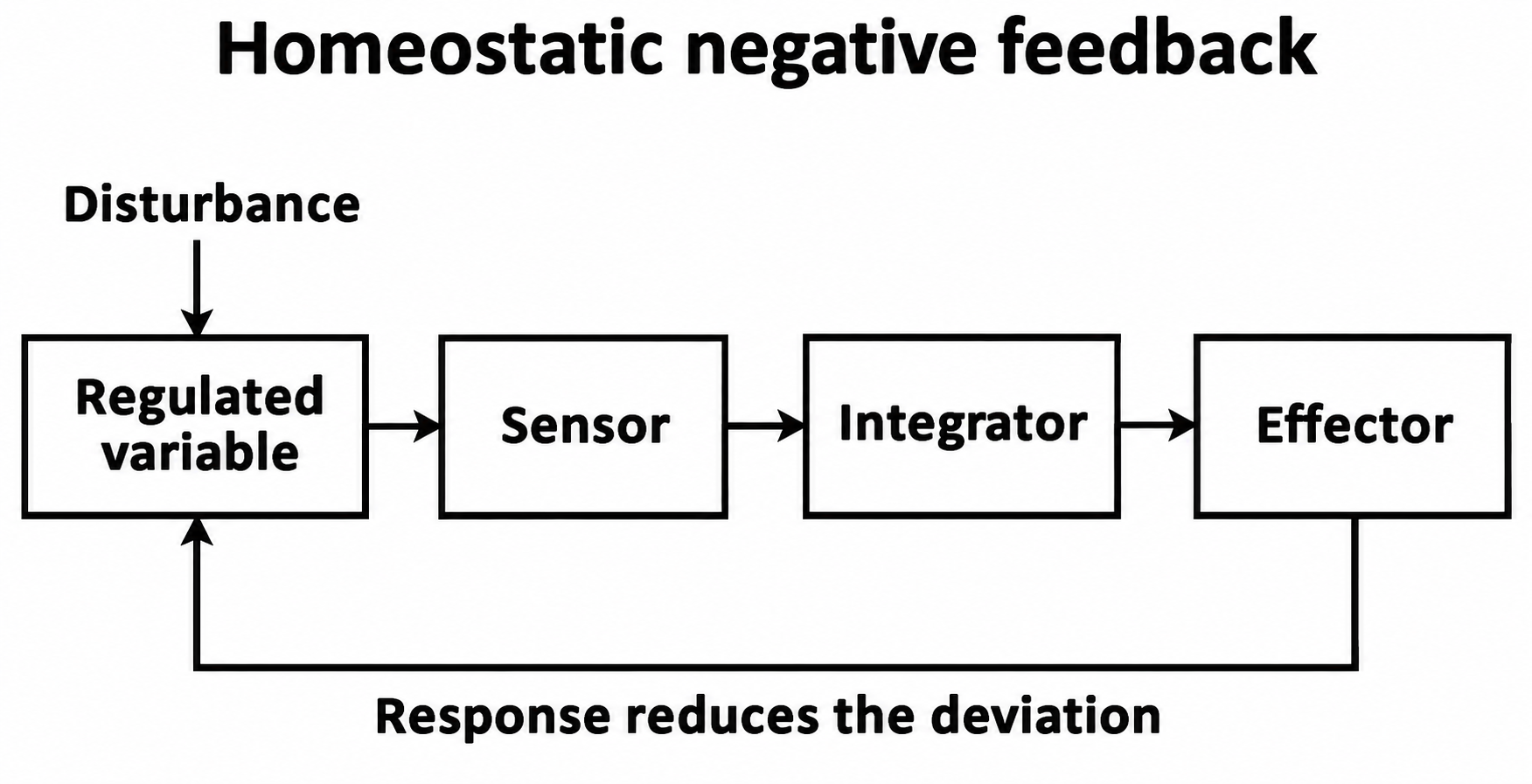
Most homeostatic control depends on negative feedback. Sensors detect a disturbance, and the body responds in a direction that reduces it. As the regulated variable returns toward its usual range, the stimulus for that response decreases.
The arterial baroreflex provides a familiar example. A fall in arterial pressure reduces stretch of baroreceptors in the carotid sinus and aortic arch. Their firing rate falls, changing the signals reaching the brainstem. Sympathetic activity increases, while parasympathetic activity decreases. Heart rate and contractility increase. Arteriolar constriction raises vascular resistance, while venous constriction can move blood toward the heart. Together, these responses help restore arterial pressure.
The responses develop over different timescales. Neural reflexes can change cardiovascular function within seconds. Some hormonal responses begin rapidly; others develop over minutes to hours. Their effects can persist after the initial stimulus changes. Renal retention of sodium and water changes body fluid volume more slowly and can continue over much longer periods.
Compensation preserves function without necessarily correcting the underlying problem.
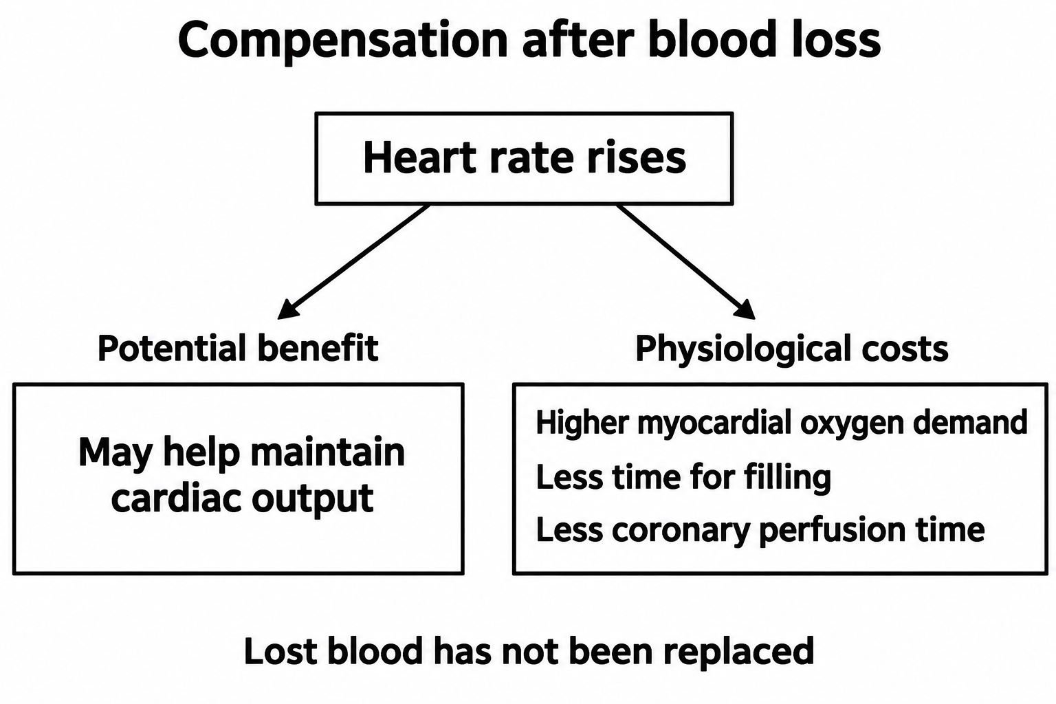
Consider acute blood loss. Tachycardia and vasoconstriction can help maintain arterial pressure. They do not replace the lost blood. If bleeding continues, the compensatory response may become insufficient.
The same responses can create new problems. A faster heart rate can help maintain cardiac output because the heart ejects blood more often. It also increases myocardial oxygen demand and shortens diastole, reducing the time available for ventricular filling and much of left ventricular coronary perfusion. Arteriolar constriction supports pressure but may reduce flow to tissues supplied by those vessels.
Metabolic acidosis produces another example. Increased ventilation removes more carbon dioxide. This lowers arterial carbon dioxide tension and partly offsets the fall in pH caused by the metabolic disturbance. The respiratory muscles must work harder to maintain that ventilation. If they can no longer do so, carbon dioxide rises and pH falls further.
Interpret pH together with bicarbonate and arterial carbon dioxide tension. A low carbon dioxide tension may be helping to offset a metabolic acidosis. If pH is nearly normal despite a major fall in bicarbonate, assess for an additional respiratory alkalosis rather than assuming that compensation alone explains the result.
Positive feedback amplifies the original disturbance. Severe myocardial ischemia can impair contraction and lower cardiac output. If arterial pressure then falls, coronary perfusion may decrease. The resulting reduction in myocardial oxygen supply can worsen ischemia and further impair contraction. Treatment must interrupt the process that is sustaining this cycle.
1.3 Physiological Reserve: The Capacity to Meet Additional Demand
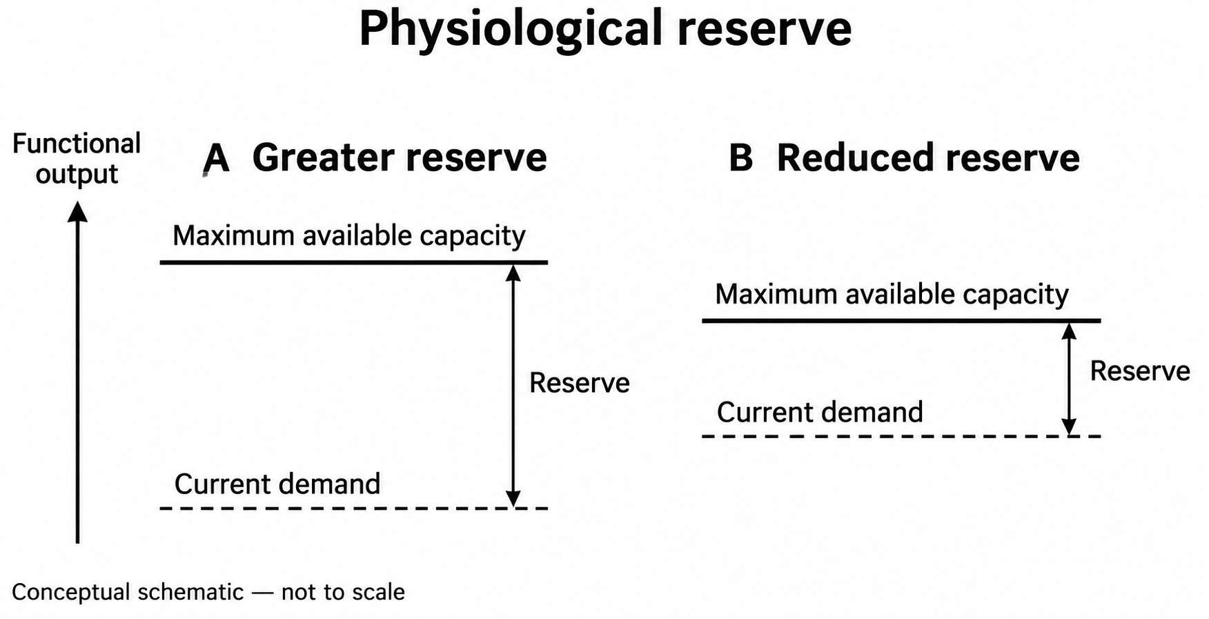
Physiological reserve is the capacity to increase function above the level currently required.
A healthy person can increase cardiac output during exercise. The kidneys can adjust electrolyte excretion when intake changes. The respiratory system can increase ventilation when carbon dioxide production rises.
These capacities are not unlimited. They also differ between organs and between patients.
A patient with severe chronic lung disease may maintain adequate gas exchange at rest. A respiratory infection adds airway obstruction, secretions, and increased metabolic demand. The additional load may exceed the patient’s remaining ventilatory capacity.
Another patient may have adequate cardiac output at rest despite severe ventricular dysfunction. Fever increases metabolic demand. Anemia reduces the oxygen carried in each unit of blood, so maintaining oxygen delivery may require more flow. A tachyarrhythmia can shorten filling time and reduce effective ejection. A ventricle that meets resting needs may be unable to meet these additional demands.
Reserve therefore depends on both available capacity and current demand. A patient can become vulnerable because capacity has fallen, because demand has increased, or because both changes have occurred together.
Reserve cannot usually be measured with one resting result. Serum creatinine mainly helps estimate current filtration and also depends on creatinine production. It does not directly measure the kidney’s ability to increase filtration or withstand a new injury. Ejection fraction describes the proportion of end-diastolic ventricular volume ejected with each beat. A preserved fraction does not show how much extra flow the circulation can provide during stress.
Premorbid function helps place these measurements in context. Relevant information includes whether the patient could walk independently, climb stairs, manage daily activities, and recover from recent illnesses.
Frailty describes a broader vulnerability to stress. It overlaps with reduced physiological reserve, although the two terms are not interchangeable. The VIP2 study examined 3,920 acutely admitted ICU patients aged 80 years or older. Premorbid frailty was associated with short-term mortality after adjustment for other factors. As an observational study, it established an association rather than a causal relationship. Its population also limits direct extrapolation to younger patients. 2
Frailty should inform a comprehensive assessment. It cannot determine an individual patient’s outcome or the appropriateness of treatment on its own.
1.4 Critical Illness: When Demand Exceeds the Capacity to Respond
Critical illness develops when an acute disturbance threatens vital function. The disturbance may overwhelm otherwise healthy organs. It may also exceed the smaller reserve of organs affected by chronic disease.
The initial problem can take many forms. Hemorrhage reduces circulating blood volume. Pneumonia impairs gas exchange. Pulmonary embolism increases the load on the right ventricle. Infection can provoke a host response that injures organs.
Sepsis also involves responses that damage the patient. The Sepsis-3 consensus defines sepsis as life-threatening organ dysfunction caused by a dysregulated host response to infection. Immune, endothelial, and circulatory disturbances can impair organ function while the body is responding to the infection. The host response can therefore contribute to injury as well as defense. 3
At the bedside, deterioration often reflects a combination of the original disease, the compensatory response, and secondary injury.
For example, a patient with severe pneumonia may initially maintain ventilation by breathing harder. As the lungs become less compliant, the respiratory muscles must generate more pressure to deliver each breath. If muscle capacity becomes insufficient, effective alveolar ventilation falls and arterial carbon dioxide tension rises. A carbon dioxide tension that moves from low toward the normal range can therefore signal deterioration when the patient remains under marked respiratory stress.
The transition to organ failure may be abrupt. Compensation can conceal progressive loss of capacity until little reserve remains. A small additional insult may then produce a large clinical change.
Follow how the patient changes over time. Increasing work of breathing, worsening mentation, or a rising requirement for cardiovascular support can reveal deterioration before arterial pressure or blood gas values cross a familiar threshold.
1.5 The Body Functions as an Interdependent System
Organ function depends on communication and exchange between organs. The lungs regulate gas exchange. The circulation transports gases and substrates. The kidneys regulate water, electrolytes, and acid excretion. The nervous and endocrine systems coordinate responses to changing conditions.
Failure in one organ can increase the workload of another.
Metabolic acidosis requires the lungs to remove more carbon dioxide to limit the fall in pH. If lung disease or muscle weakness prevents the necessary increase in ventilation, pH falls further. The interaction also works in the other direction. Alveolar hypoxia can constrict pulmonary vessels and raise the resistance against which the right ventricle ejects. Severe lung disease can therefore increase right ventricular work while impairing oxygenation.
Treatment also acts across organs. Positive-pressure ventilation can take over part of the respiratory muscles’ work. The accompanying rise in intrathoracic pressure can reduce venous return, while lung inflation can increase right ventricular afterload. Intravenous fluid may increase cardiac output when the ventricles respond to additional filling. It can also raise cardiac filling pressures. Higher pressure transmitted to pulmonary or systemic veins can worsen congestion, even when output increases.
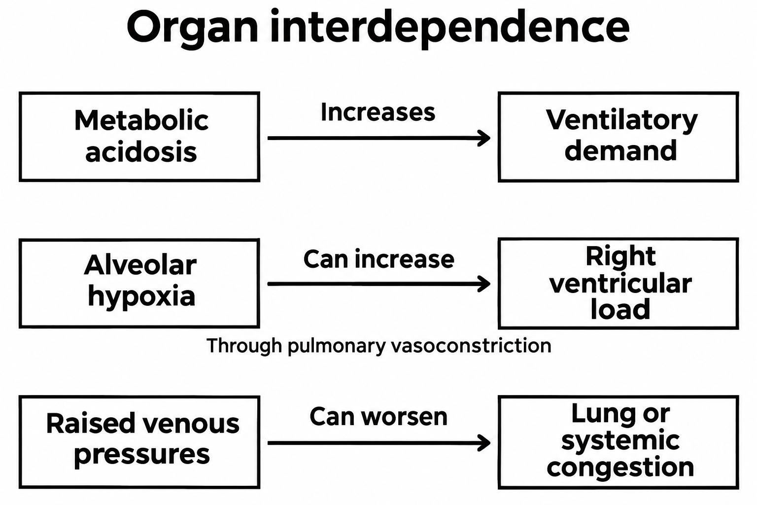
These interactions explain why a treatment can improve one variable while creating another problem.
Blood must reach the microcirculation before oxygen can pass into tissue. A systemic arterial pressure measurement does not show which small vessels are carrying blood or whether flow is evenly distributed. Obstructed or poorly perfused microvessels can leave some tissue regions undersupplied despite an acceptable systemic pressure.
In a human observational study, De Backer and colleagues demonstrated abnormal small-vessel perfusion in patients with sepsis. These abnormalities were more pronounced in patients with worse outcomes. The findings support the importance of microvascular dysfunction, but they do not establish that a particular treatment directed at the microcirculation improves survival. 4
Restoring systemic pressure is therefore an important component of circulatory support. It does not, by itself, demonstrate that tissue perfusion has recovered.
1.6 Interpreting Measurements: A Signal Is Not a Mechanism
A physiological measurement provides information about a process. It rarely identifies the entire process.
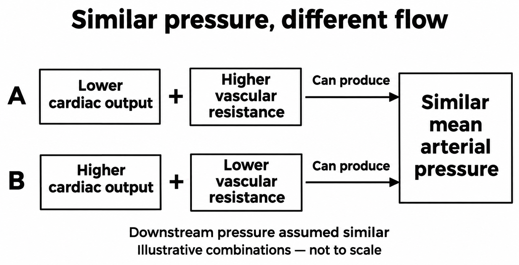
Arterial pressure depends on both cardiac output and the resistance to blood flow. Low output combined with high resistance can produce a pressure similar to that produced by high output and low resistance. The same pressure can therefore accompany very different causes of circulatory failure.
Lactate requires the same care in interpretation. Its concentration rises when production exceeds removal. Inadequate oxygen supply can increase lactate production, but it is not the only explanation. Rapid glycolysis can produce more lactate even when oxygen is available. Reduced removal by the liver and other tissues can raise the concentration further.
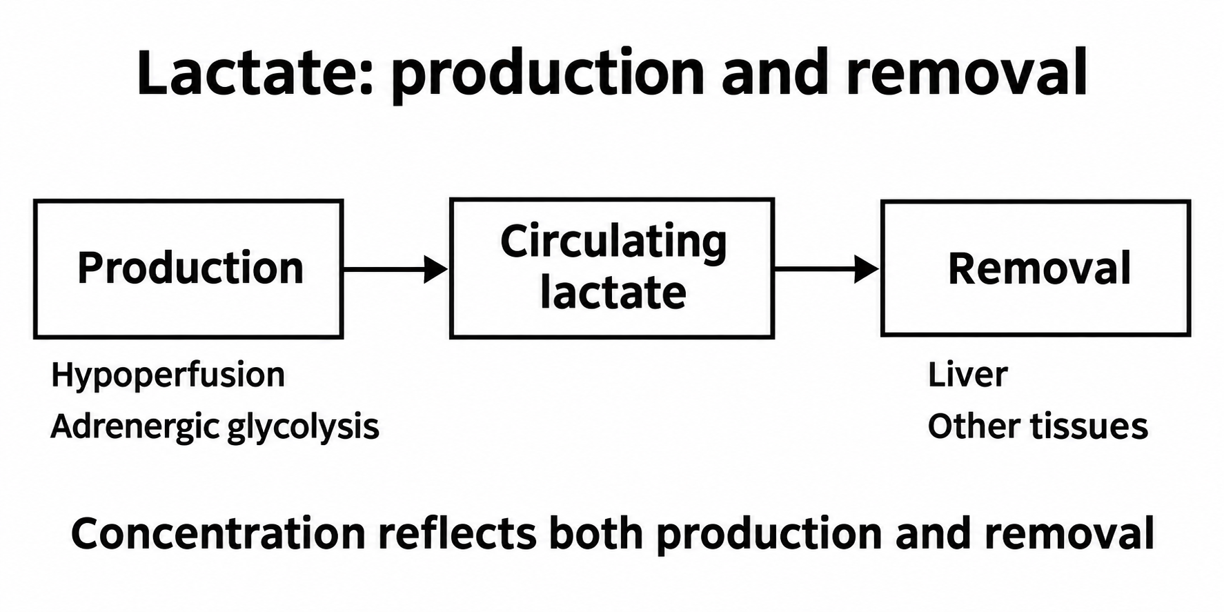
James and colleagues demonstrated epinephrine-stimulated aerobic lactate production in isolated rat skeletal muscle. This experimental mechanism illustrates why hyperlactatemia need not mean oxygen debt; it does not determine the cause in an individual patient. 5
An elevated lactate remains clinically important. It should prompt assessment of perfusion, disease severity, and possible causes. It does not automatically identify fluid administration as the appropriate treatment.
Low urine output also has several possible causes. Reduced renal perfusion can lower filtration. High renal venous pressure can impair drainage and reduce the pressure difference across the kidney. Established kidney injury can impair filtration or tubular function, and obstruction can prevent urine from leaving. Hormonal sodium and water retention can reduce urine output without establishing a volume deficit. The next step depends on which process is occurring.
A useful approach is to compare several observations that reflect different aspects of function. In circulatory failure, these may include arterial pressure, capillary refill, skin temperature, mental status, urine output, and lactate. Each has limitations. Agreement between them strengthens an interpretation. Disagreement should prompt reassessment.
ANDROMEDA-SHOCK-2 tested a protocol that combined capillary refill assessment with hemodynamic evaluation. Its primary analysis included 1,467 patients with early septic shock. The main outcome compared death first, then the duration of vital support, then hospital stay through day 28. The protocol improved this combined outcome, mainly through shorter vital support. Mortality at 28 days was similar. The result supports the complete strategy; it does not isolate the benefit of measuring capillary refill. 6
1.7 Supporting Physiology Without Chasing Normality
A reference range describes measurements in a reference population. It does not automatically define the best treatment target during critical illness.
The distinction is important because an intervention has effects beyond the variable it changes.
The NICE-SUGAR trial provides a clear example. Intensive glucose control targeted 81–108 mg/dL. Conventional control used a target of 180 mg/dL or less. Ninety-day mortality was higher with intensive control: 27.5% compared with 24.9%. Severe hypoglycemia was also more frequent. Bringing glucose closer to a healthy fasting range did not improve outcomes in this population. 7
Blood-pressure targets illustrate a related trade-off. In SEPSISPAM, 776 patients with septic shock were assigned to a mean arterial pressure target of either 80–85 mm Hg or 65–70 mm Hg. The higher target did not significantly reduce mortality. New atrial fibrillation was more frequent in the higher-target group. Patients with chronic hypertension required less renal replacement therapy at the higher target, but this subgroup finding was not accompanied by a mortality benefit. 8
In OPTPRESS, targeting a mean arterial pressure of 80–85 mm Hg increased 90-day mortality compared with 65–70 mm Hg in older Japanese patients with septic shock. This later trial adds a reason not to use age or chronic hypertension alone to justify a high pressure target. 10
These trials do not make physiological reasoning less important. They show why physiological reasoning must be tested against patient outcomes.
Before changing treatment, specify what should improve. If low arterial pressure is limiting organ perfusion, a pressure increase should improve findings consistent with that problem. Before giving fluid, establish whether more flow is needed and whether additional filling is likely to increase it. Also identify the findings that would prompt treatment to stop, such as worsening pulmonary edema or arrhythmia.
The response should then be reassessed. A treatment that changes the target measurement without producing the expected physiological benefit may require reconsideration.
1.8 Time, Treatment Dependence, and Recovery
Physiology changes throughout an illness. The same measurement can have a different meaning at different times.
A stable mean arterial pressure with a falling norepinephrine requirement suggests a different trajectory from the same pressure with a rapidly rising requirement. The observed pressure may be identical. The patient’s dependence on treatment is changing.
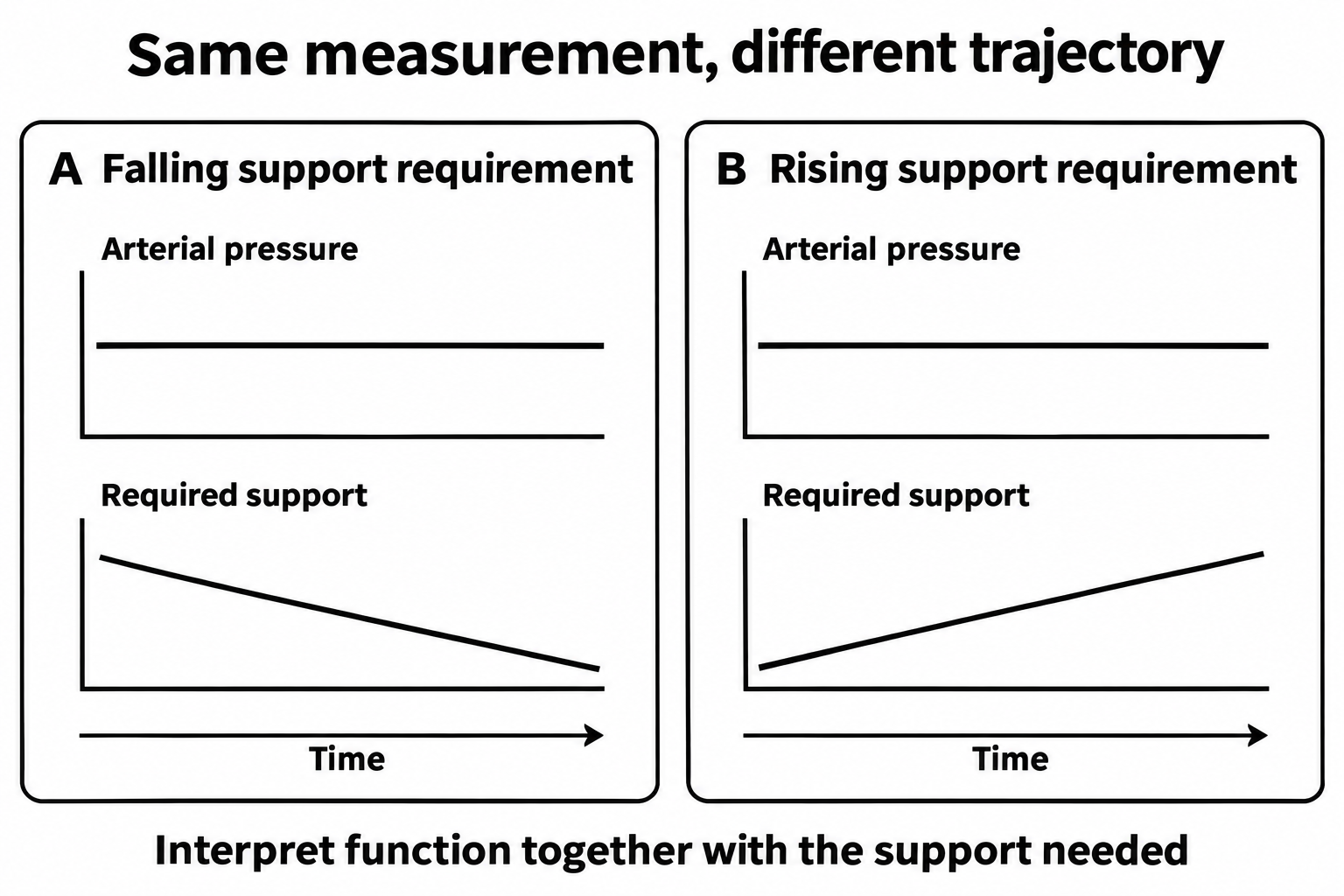
This principle also applies to respiratory support. An unchanged oxygen saturation may represent improvement if the required inspired oxygen concentration is falling. It may represent deterioration if substantially more support is needed to maintain it.
External support can preserve function while the underlying disease is treated. It does not necessarily restore the patient’s capacity to function independently. A ventilator can maintain ventilation in a patient whose respiratory muscles remain weak. Kidney replacement therapy can control potassium and acid–base balance while native kidney function remains inadequate.
Recovery must therefore be assessed through both organ function and the amount of support required.
Recovery also extends beyond the resolution of acute organ failure. The BRAIN-ICU study found persistent cognitive impairment among survivors of respiratory failure or shock. Longer delirium duration was associated with worse subsequent cognitive performance. Because this was an observational study, the association does not establish that delirium alone caused the impairment. 9
A patient may leave the ICU with satisfactory vital signs yet have substantial physical or cognitive limitations. Restoration of independence requires a broader view of recovery than normalization of bedside measurements.
1.9 Applying the Principles at the Bedside
Consider an illustrative patient with pneumonia and septic shock.
The patient is 74 years old and has chronic heart failure and chronic kidney disease. After initial treatment, the mean arterial pressure is 70 mm Hg on norepinephrine. Lactate has fallen from 5.0 to 3.8 mmol/L. Urine output remains low. The oxygen saturation is 94% on an inspired oxygen fraction of 0.60. The patient is therefore breathing a mixture containing 60% oxygen to maintain that saturation.
These observations are insufficient to establish that resuscitation is complete. They also do not establish that additional fluid is needed.
First establish whether the patient is improving or deteriorating. Determine whether the norepinephrine dose is falling or rising. Reassess mental status and capillary refill. Check the effort required to breathe and whether more or less oxygen is needed to maintain the same saturation.
The next task is to identify the likely mechanism of any persistent dysfunction. Low urine output could reflect reduced renal perfusion, kidney injury, or congestion. A persistently elevated lactate could reflect ongoing hypoperfusion, increased production through other pathways, impaired removal, or several mechanisms together.
The patient’s baseline disease matters. Heart failure may limit the ability to increase cardiac output. Chronic kidney disease may reduce tolerance of further injury. Neither diagnosis, by itself, predicts the response to fluid.
If additional fluid is being considered, two separate issues require assessment. The first is whether an increase in preload could produce a useful increase in cardiac output. The second is whether the patient could tolerate the associated increase in filling pressures. A patient may be fluid responsive and still be vulnerable to pulmonary edema or venous congestion.
The response to treatment should address the original physiological concern. A rise in arterial pressure alone may not establish whether tissue perfusion has improved. Conversely, a persistently abnormal creatinine concentration may not indicate treatment failure if perfusion is improving and kidney recovery is delayed.
Connect each abnormal finding to a possible mechanism. Decide whether the patient can sustain the current work of breathing and circulatory demands. Account for the support already needed to maintain function. Give each intervention a clear purpose, then check whether it produces the expected benefit and whether it causes harm. Use the response to revise the next decision.
References
1. Cannon WB. Organization for physiological homeostasis. Physiological Reviews. 1929;9(3):399–431. doi:10.1152/physrev.1929.9.3.399.
2. Guidet B, de Lange DW, Boumendil A, et al. The contribution of frailty, cognition, activity of daily life and comorbidities on outcome in acutely admitted patients over 80 years in European ICUs: the VIP2 study. Intensive Care Medicine. 2020;46:57–69. PubMed.
3. Singer M, Deutschman CS, Seymour CW, et al. The Third International Consensus Definitions for Sepsis and Septic Shock (Sepsis-3). JAMA. 2016;315(8):801–810. doi:10.1001/jama.2016.0287.
4. De Backer D, Creteur J, Preiser JC, Dubois MJ, Vincent JL. Microvascular blood flow is altered in patients with sepsis. American Journal of Respiratory and Critical Care Medicine. 2002;166(1):98–104. doi:10.1164/rccm.200109-016OC.
5. James JH, Wagner KR, King JK, et al. Stimulation of both aerobic glycolysis and Na+-K+-ATPase activity in skeletal muscle by epinephrine or amylin. Am J Physiol. 1999;277:E176–E186. Original experimental study.
6. The ANDROMEDA-SHOCK-2 Investigators. Personalized hemodynamic resuscitation targeting capillary refill time in early septic shock: the ANDROMEDA-SHOCK-2 randomized clinical trial. JAMA. 2025;334(22):1988–1999. doi:10.1001/jama.2025.20402.
7. NICE-SUGAR Study Investigators. Intensive versus conventional glucose control in critically ill patients. New England Journal of Medicine. 2009;360:1283–1297. doi:10.1056/NEJMoa0810625.
8. Asfar P, Meziani F, Hamel JF, et al. High versus low blood-pressure target in patients with septic shock. New England Journal of Medicine. 2014;370:1583–1593. doi:10.1056/NEJMoa1312173.
9. Pandharipande PP, Girard TD, Jackson JC, et al. Long-term cognitive impairment after critical illness. New England Journal of Medicine. 2013;369:1306–1316. doi:10.1056/NEJMoa1301372.
10. Endo A, Yamakawa K, Tagami T, et al.; OPTPRESS trial investigators. Efficacy of targeting high mean arterial pressure for older patients with septic shock (OPTPRESS): a multicentre, pragmatic, open-label, randomised controlled trial. Intensive Care Medicine. 2025;51:883–892. doi:10.1007/s00134-025-07910-4.